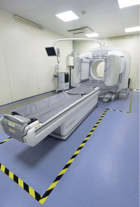
Introduction and Function Description of GE Spect CT
1、 Equipment Introduction
GE Spect CT, The full name is Single Photon Emission Computed Tomography/Computed Tomography (SPECT/CT), which is currently one of the advanced medical imaging devices in the field of nuclear medicine. This device combines single photon emission computed tomography (SPECT) and computed tomography (CT) technologies, achieving a perfect combination of functional metabolic imaging and anatomical structure imaging.
The GE Spect CT series includes multiple models, such as Discovery NM/CT 670 Pro, NM/CT 860, etc. These devices play an important role in clinical diagnosis with their high sensitivity and specificity. They use advanced detectors and image processing technology, which can utilize the special physiological functions of radioactive tracers in the human body to accurately locate and qualitatively diagnose the lesion site through high-precision imaging technology.
2、 Function Description
Whole body skeletal imaging
Early diagnosis of bone metastases: SPECT/CT bone imaging can detect bone metastases earlier than ordinary X-ray or CT examination. For breast cancer, lung cancer, prostate cancer and other diseases prone to bone metastases, metastasis can be found 3-6 months or even earlier.
The diagnosis of bone tumors and osteonecrosis: helps to determine the extent of involvement of primary bone tumors, monitor the treatment effect of bone tumors, and detect early lesions such as osteonecrosis.
Diagnosis of thyroid and parathyroid diseases
Diagnosis of thyroid diseases: Used for auxiliary diagnosis of hyperthyroidism and thyroiditis, evaluating thyroid size, distinguishing benign and malignant thyroid nodules, and locating differentiated thyroid cancer metastases.
Diagnosis of Parathyroid Diseases: Used for the diagnosis and localization of parathyroid adenomas, and to evaluate the parathyroid status of patients with hyperparathyroidism.
Renal function testing
Dynamic renal function test: evaluate the glomerular filtration rate, monitor the function of transplanted kidney, identify renal hypertension, and observe the impact of diabetes on renal function.
Upper urinary tract patency assessment: Diagnose urinary tract obstruction, evaluate renal artery disease and bilateral renal blood supply.
Cardiovascular system diagnosis
Diagnosis of myocardial ischemia: Evaluate the extent of coronary artery disease, assess myocardial cell viability, evaluate the prognosis of myocardial infarction, and observe the improvement of myocardial ischemia after cardiac bypass surgery and interventional therapy.
Cardiomyopathy auxiliary diagnosis: By using myocardial perfusion imaging, the blood supply of the myocardium can be understood to assist in the diagnosis of cardiomyopathy.
Diagnosis of neurological disorders
Localization of epileptic lesions: Through cerebral blood flow perfusion imaging, epileptic lesions can be located to provide a basis for surgical treatment.
Early diagnosis of cerebral infarction: In the early stage of cerebral infarction, abnormalities can be displayed when there are no obvious structural changes in the affected area, which is helpful for early diagnosis and treatment.
Digestive system diagnosis
Assessment of gallbladder contraction function: Understanding the contraction function of the gallbladder to assist in the diagnosis of gallbladder diseases.
Analysis of gastrointestinal bleeding: Diagnosis of the location and cause of gastrointestinal bleeding using radiopharmaceutical labeled imaging agents.
Other applications
Salivary gland imaging: evaluates the uptake and secretion function of salivary glands and the patency of ducts, and diagnoses diseases such as Sjogren's syndrome.
Lymphatic imaging: Evaluating lymphatic reflux and assisting in the diagnosis of lymphatic system diseases.
3、 Technical features
Fusion imaging technology: SPECT/CT combines the functional metabolic imaging of SPECT with the anatomical structure imaging of CT, completing both nuclear medicine SPECT and radiation CT examinations in one examination, which not only understands the functional status of lesions, but also achieves precise localization.
High sensitivity and specificity: Using low-dose, short half-life radioactive nuclide drugs for tracing can determine disease characteristics at the molecular and metabolic levels, improving the sensitivity and specificity of disease diagnosis.
Advanced detector technology: Some high-end models from GE use CZT (cadmium zinc telluride) detectors, which have high sensitivity and resolution and can achieve high-definition imaging.
Intelligent image processing: Equipped with advanced image processing software, it can automatically perform image fusion, reconstruction, and analysis, improving diagnostic efficiency and accuracy.
As an advanced equipment in the field of nuclear medicine, GE Spect CT plays an important role in clinical diagnosis with its fusion imaging technology, high sensitivity and specificity, advanced detector technology, and intelligent image processing capabilities. It can achieve early diagnosis and precise positioning of various diseases, providing important basis for the treatment of patients.

